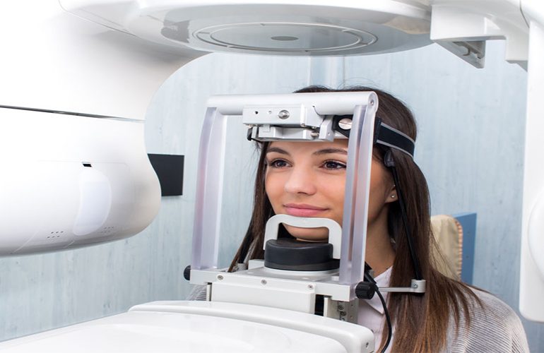
When you receive a tomography report, the language you encounter is often difficult to understand. Expressions such as “radiolucent area,” “cortical continuity” and “crestal bone level” may seem worrying to someone unfamiliar with medical terminology. However, most of these terms are descriptive; that is, they describe what is seen and do not declare a disease on their own. As DijiDent Oral and Dental Health Polyclinic, we offer our patients in Türkiye this guide to help them understand their own health records.
First, the Basic Distinction: Image and Interpretation
An imaging record consists of two parts. The first is the raw data, that is, all the slices produced by the device. The second is the interpretation that emerges from the evaluation of this data by a specialist. The report is the written form of the second part. The points emphasized by two dentists looking at the same image may differ; this does not necessarily mean a contradiction and may stem from differences in clinical priorities.
Frequently Encountered Terms and Their Meanings
Radiolucent and Radiopaque
Areas that appear dark in the image are described as radiolucent, and areas that appear light and bright as radiopaque. Dense structures such as bone and tooth tissue appear light; cavities, soft tissues or areas with bone loss appear dark. The expression “radiolucent area” on its own is not a bad finding; the sinus cavity also appears radiolucent. What matters is where this area is and what borders it has.
Cortical Bone and Continuity
The hard layer on the outer surface of the bone is called cortical bone. The expression “cortical continuity preserved” indicates that the integrity of this outer wall has not been disrupted and is generally a positive finding.
Crestal Bone Level
This refers to the uppermost border of the bone surrounding the teeth. If this level has moved down along the root, it suggests bone loss due to gum disease and requires periodontal evaluation.
Periapical Lesion
This describes the dark area at the root tip, usually developing due to canal-related infection. It is a finding that requires endodontic evaluation.
Mandibular Canal and Maxillary Sinus
These refer to the canal through which the nerve passes in the lower jaw and the air cavity in the upper jaw. Stating the distance to these structures in the report is important for surgical planning.
Artifact
This describes the glare and streaking that a metal filling, crown or implant creates in the image. It is not a real pathology but an effect originating from the imaging.
What Should You Pay Attention to in the Report?
- Date: How current the image is directly affects the validity of the interpretation.
- Imaged area: The report covers only the region scanned; it gives no information about structures outside it.
- Measurement values: If bone height and width are stated in millimeters, they can be used for planning.
- Recommendation section: The expression “clinical correlation is recommended” is frequently used; it indicates that the finding should be evaluated together with the examination.
The Same Finding Can Carry Different Meanings
The seriousness of an expression in the report changes depending on its context. A small dark area seen at the root tip may belong to a region treated years ago that is healing, or it may be a sign of an active infection. To make this distinction, a comparison with your previous images is needed. This is why we care about you keeping your old records: change over time carries much more information than a single image can provide.
The Report Is Not a Diagnosis
This is the point we most want to emphasize. An imaging report gains meaning when combined with your complaints and clinical examination findings. Searching an expression from the report on the internet and focusing on the worst possibility leads to unnecessary anxiety. Asking your dentist about every line you do not understand is the right way; at our clinic, we expect these questions.
Frequently Asked Questions
This expression is not a definitive diagnosis; it indicates that additional evaluation or follow-up is needed. Clarify the next step by talking to your dentist.
The images are generated in a short time. The time for the written evaluation varies depending on the scope of the case and the clinical workflow.
Of course. Getting a second opinion is your right; for this, it is recommended that you also share the image file.
Understanding the general concepts makes the process easier; however, a clinical decision requires a dentist’s evaluation.
If there has been treatment, extraction or change in your mouth, a current evaluation may be needed. The decision belongs to your dentist.
Understanding your own health record is your right.
Let us examine your tomography or report together and explain in plain language what the findings mean. Contact us to book an appointment; let us carry out the process transparently at DijiDent Oral and Dental Health Polyclinic.
This content is for general information purposes and does not replace a diagnosis. Report findings are evaluated by your dentist together with the clinical examination.