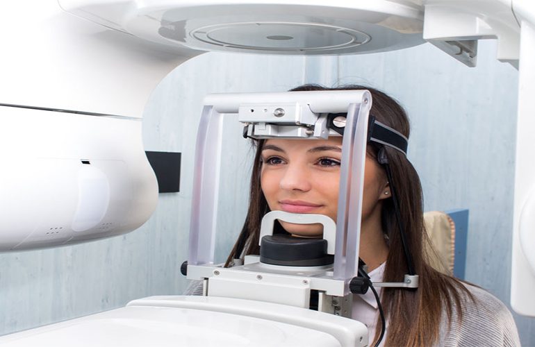
When you go to your dentist, you have probably heard the sentence “let’s take a film first.” In some cases, however, your dentist is not satisfied with that alone and asks for three-dimensional imaging. At this point many patients rightly ask: why is a tomography needed when I already have a film? The difference between 3D tomography and panoramic X-ray actually comes from the fact that they look for answers to two different questions. As DijiDent Oral and Dental Health Polyclinic, in the imaging services we offer in Türkiye, we consider clearly explaining to our patients which examination is requested and why to be part of the treatment.
What Does a Panoramic X-Ray Show?
A panoramic X-ray offers a flattened summary of your entire jaw in a single image. As the device rotates around your head, it gathers all the teeth in the upper and lower jaws, the general structure of the jawbone and part of the sinus regions into a single frame. This method provides a fast and effective starting point for evaluating the number of teeth, the presence of impacted teeth, large cavities, areas suspected of cysts and the general bone level.
The greatest advantage of the panoramic film is its practicality. It is taken in a short time, works with a low dose and provides sufficient information in most routine check-ups. However, there is an important limit here: a panoramic X-ray is a two-dimensional image. In other words, it carries no depth information.
The Limit of a Two-Dimensional Image
When you transfer a three-dimensional structure onto a two-dimensional plane, structures lying behind one another overlap. On a panoramic film, a tooth root and the mandibular nerve may appear intertwined, whereas in reality there may be a safe distance of a few millimeters between them. Or the opposite may be true: two structures that appear far apart on the film may actually be adjacent. In addition, magnification ratios and positioning errors in panoramic images affect measurement accuracy. For this reason, it is not considered sufficient on its own in situations that require millimetric decisions.
What Does 3D Dental Tomography Do Differently?
Cone beam computed tomography (CBCT) creates a volumetric model of the region. Your dentist can slice this model in any direction, rotate it and take true-to-size measurements. In practice, this means the following:
- Bone thickness can be measured: In the area where the implant will be placed, not only the height of the bone but also its width in the lip-palate direction can be seen.
- Anatomical relationships become clear: The relationship with the mandibular nerve, the sinus floor and neighboring tooth roots is examined in three planes.
- Hidden pathologies may be revealed: A small lesion hidden behind a tooth root on a two-dimensional film may be noticed in a cross-sectional image.
- Canal anatomy can be seen: In recurring infections, an overlooked additional root canal can be detected.
- Surgery is planned in advance: When the angle and root structure of an impacted tooth are known, procedure time and complication risk are better managed.
Which One Is Needed in Which Case?
These two methods are not rivals of each other; they are complementary. The general approach can be summarized as follows: for routine examinations, caries screening and general evaluation, a panoramic X-ray is often sufficient. On the other hand, three-dimensional imaging makes a significant contribution in implant planning, impacted tooth surgery, root canal retreatment, cyst-lesion evaluation, jaw joint complaints and post-trauma examinations.
Our principle at DijiDent is clear: imaging is requested if it will answer a clinical question. Instead of applying tomography to everyone as a routine procedure, we recommend it in cases where it will provide information that directly affects your treatment decision. Our dentists also explain this rationale to you verbally; we do not think it is right for you to proceed without knowing what we are asking for and why.
Is My Old Film Useful?
The images you bring with you are definitely valuable; they allow comparison, especially in terms of showing the past situation. However, the date, quality and the area covered by the image are decisive. A film taken years ago may not reflect the current bone level. For this reason, be sure to bring your existing film; let your dentist decide after the examination whether additional imaging is needed.
Frequently Asked Questions
No. The panoramic X-ray retains its value as a fast, low-dose, general screening tool. Tomography comes into play when a specific question requires a detailed answer.
3D tomography generally uses a higher dose than a panoramic X-ray and a significantly lower dose than medical computed tomography. Reducing the imaging field is an important factor in lowering the dose. Exact values vary depending on the device and the imaging protocol.
It is possible if there is a clinical need; however, the aim is to avoid unnecessary repetition. Your dentist determines whether an additional examination is needed after reviewing your existing images.
Yes. Images can be shared as digital files; they can be used to obtain a second opinion or for follow-up at another center.
Both types of imaging are painless. You are only asked to remain still for a few seconds.
The right examination also prevents unnecessary treatment.
Contact us to book an appointment to find out whether your existing films are sufficient and to build your treatment plan on a solid diagnosis. Let our dentists at DijiDent Oral and Dental Health Polyclinic evaluate your images together with you.
This content is for general information purposes and does not replace an examination by a dentist. The imaging decision is made based on clinical evaluation.