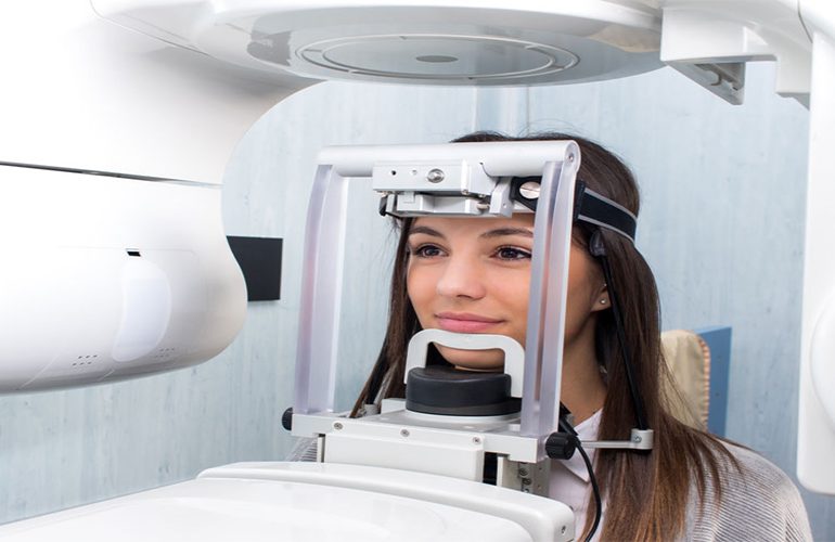
Wisdom teeth remain impacted or only partially erupt when they cannot find enough space in the jaw. This condition often progresses silently, until recurring gum inflammation, caries in the neighboring molar or a persistent pain appears. When the surgical decision is being made, the most critical question is this: how close is the relationship between the roots of the tooth and the mandibular nerve? Impacted tooth tomography answers this question without leaving it to guesswork. As DijiDent Oral and Dental Health Polyclinic, in the surgical planning we carry out in Türkiye, we consider seeing the risk in advance to be a prerequisite for safety.
Why Is a Panoramic Film Not Always Sufficient?
A panoramic X-ray shows the presence and approximate position of the impacted tooth. However, because it is a two-dimensional image, it carries no depth information. On the film, the root of the tooth and the nerve canal may appear to overlap; this image alone does not mean that the two are in contact. The canal may be on the tongue side of the root or on the cheek side. These three possibilities completely change the surgical approach.
In the dental literature, three-dimensional imaging is recommended when certain risk signs are seen on a panoramic film. Darkening of the root over the canal, deviation in the course of the canal or the loss of the canal borders are among these signs. At DijiDent, when we detect these findings, we recommend cross-sectional imaging instead of proceeding blindly.
What Does Tomography Add to the Surgical Plan?
- Nerve proximity becomes clear: The true distance and directional relationship between the root tips and the mandibular nerve can be measured.
- Root morphology is seen: Curved, divergent or interlocked roots are known in advance; the technique is chosen accordingly.
- The angle of the tooth is determined: Horizontal, angled or vertical position determines from which direction the tooth will be approached.
- The effect on the neighboring tooth is understood: Root resorption or the onset of caries in the adjacent molar can be detected.
- The sinus relationship in the upper jaw is seen: In upper wisdom teeth, the proximity of the root to the sinus cavity is evaluated.
- The duration becomes predictable: When the degree of difficulty is known, the procedure time and recovery expectations are communicated to you more realistically.
Why Is It Important to Know the Degree of Difficulty in Advance?
How long the surgical procedure will take, whether bone will need to be removed and whether the tooth will be removed by sectioning are directly related to knowing the root structure in advance. This information is valuable not only for the dentist but also for you: you can plan the duration of the procedure, what to expect during recovery and your work or school schedule accordingly. As uncertainty decreases, clinical safety increases and the patient’s sense of control over the process becomes stronger.
Should Every Impacted Tooth Be Extracted?
No. This is one of the topics our patients most often have misconceptions about. Some teeth that remain completely within the bone, do not press on the neighboring tooth and do not cause any pathology can be monitored regularly. On the other hand, partially erupted teeth present a different picture: food and bacteria accumulate under the gum flap, cannot be cleaned and pave the way for recurring inflammation (pericoronitis). The decision is made by evaluating the clinical examination, imaging findings and your complaints together.
Pre-Surgical Preparation and Information
When imaging is completed, your dentist shares the findings with you on screen. Seeing where the tooth is located, which structures it is adjacent to and the planned approach while listening significantly reduces anxiety about the procedure. At this stage, you need to fully share the medications you use, especially blood thinners, and your chronic diseases. Do not stop your medications on your own decision; the necessary adjustment is planned together with your relevant specialist physician.
Frequently Asked Questions
No. If there is no risk sign on the panoramic film and the tooth is in an easily accessible position, additional imaging may not be needed. Tomography is requested in cases where it will provide information that changes the decision.
Proximity alone does not prevent extraction; it changes the approach. In some cases, alternative techniques in which only the crown part is removed instead of the entire tooth can be considered. Suitability is individual.
Temporary changes in sensation can be seen in some cases and usually subside over time. Permanent cases are rare. Evaluating this risk in advance is one of the most important contributions of three-dimensional imaging.
Swelling and mild discomfort in the first days are normal. In most cases, social recovery becomes evident within a few days; the exact duration varies depending on the difficulty of the procedure and the person.
Yes. Some impacted teeth remain silent for a long time. Regular check-ups allow a problem to be noticed before it grows.
Find out whether your impacted tooth really needs to be extracted.
Contact us to book an appointment for an imaging-supported evaluation and surgical planning tailored to you. Let our dentists at DijiDent Oral and Dental Health Polyclinic talk openly with you about the risks and options.
This content is for general information purposes. The surgical decision is made by your dentist after the clinical examination and radiological evaluation.