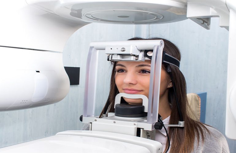
For many years, orthodontic treatment planning was carried out with the pair of panoramic film and profile X-ray; today, too, this pair is sufficient in most routine cases. However, in some situations, every step taken without knowing exactly where the tooth sits in the jaw carries risk. 3D imaging in orthodontics comes into play precisely in these selected cases. As DijiDent Oral and Dental Health Polyclinic, when carrying out orthodontic planning in Türkiye we first ask this question: will this imaging change my treatment decision? If the answer is no, we do not request the examination.
Tomography Is Not Needed in Routine Orthodontics
It is important to state this clearly: in cases of simple crowding, mild bite problems and where standard braces or clear aligner treatment is planned, three-dimensional imaging is not routinely requested. Panoramic film, cephalometric X-ray, intraoral scanning and clinical examination provide sufficient information for planning in most cases. In accordance with the radiation safety principle, if a lower-dose method answers the question, a higher-dose one is not used.
In Which Cases Does It Really Contribute?
Impacted Canine Teeth
Canines are among the teeth with the longest eruption path, and in some children they do not erupt at the expected time. The critical question is this: has the tooth remained impacted on the lip side or the palate side of the teeth? This information directly determines the path along which it will be pulled and the direction of the force to be applied. In addition, whether resorption has started at the root of the neighboring incisor can only be reliably evaluated in a cross-sectional image; this finding changes the urgency of treatment.
Extra and Developmentally Anomalous Teeth
The position of extra teeth developing outside the normal dental arch, whether they will be removed and from which direction they will be accessed become clear with a three-dimensional image.
Skeletal Discrepancy and Surgically Assisted Cases
In cases where the relationship between the jaws is significantly disturbed, where there is asymmetry or where orthognathic surgery is being considered, three-dimensional evaluation forms the basis of planning. Simulating jaw movements in a virtual environment makes it easier for the surgeon and the orthodontist to establish a joint plan.
Mini-Screw (Temporary Anchorage) Applications
Mini-screws placed in the bone to support tooth movement need to coincide with the safe space between the tooth roots. Measuring the spaces between the roots is decisive in this application.
Gum Recession and Thin Bone Structure
In some adult patients, the bone wall surrounding the teeth is thin. Evaluating in advance whether the planned movement will push the limits of this wall helps manage the risk of gum recession after treatment.
Assessing the Risk of Root Resorption
During orthodontic movement, shortening of the tooth root tips may be seen in some patients. This risk is better managed by knowing the root structure and bone boundaries before treatment. Especially in patients with thin or short root structures, the force level and treatment duration are planned accordingly. This evidence-based adjustment increases both the safety and the predictability of treatment.
Combining Imaging with Digital Impressions
At DijiDent, we do not use tomography data on its own. When the digital impression taken with an intraoral scanner is combined with the image showing the bone and tooth roots, the movement of both the crown and the root can be planned together. This is particularly important in clear aligner treatments: a tooth arrangement that looks good on screen may not be feasible if the root movements push the bone boundaries. Building the plan on these two data sets makes it possible to set realistic goals.
Frequently Asked Questions
It is not needed in routine cases. It is recommended in selected situations such as an impacted tooth, an extra tooth or significant asymmetry, and its rationale is explained to you.
Initial records are taken; if deemed necessary, a control image may be requested in the middle and at the end of treatment. The number varies by case and unnecessary repetition is avoided.
No. In many cases, the aim is to access the tooth surgically and bring it into the arch with orthodontic force. The decision depends on the position of the tooth and the condition of the neighboring roots.
It is not routinely needed. However, it can contribute in cases where complex root movements are planned or where there is gum recession.
If gum and bone health are adequate, treatment can be applied in adults. For this reason, a periodontal evaluation is recommended before orthodontics.
Build your orthodontic plan with the right records.
Contact us to book an appointment to learn the treatment approach suitable for you or your child and which records are needed. Let us carry out your orthodontic evaluation with digital data at DijiDent Oral and Dental Health Polyclinic.
This content is for general information purposes. The imaging and treatment decision is made by your dentist after the clinical examination.