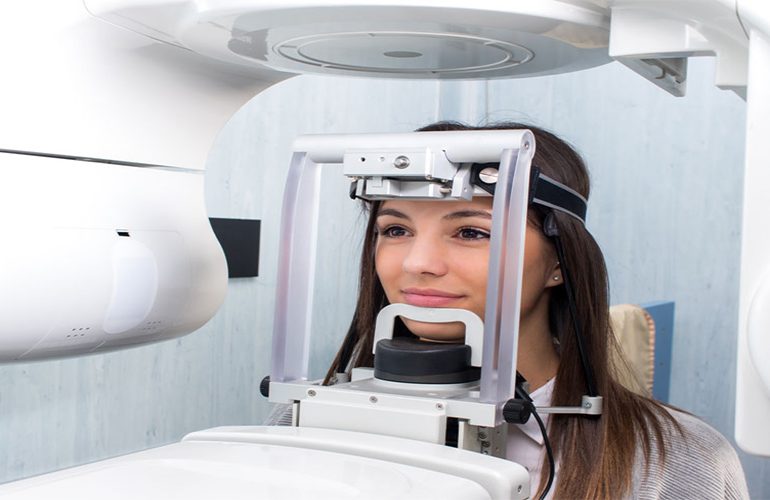
It is natural to hesitate when imaging is recommended for your child. Developing tissues are considered more sensitive to radiation than adult tissues; therefore, every examination in child patients requires more meticulous justification. However, this does not mean that imaging is a procedure to be avoided entirely. When used with the correct indication, dental tomography in children can prevent unnecessary surgery and wrong treatment decisions. As DijiDent Oral and Dental Health Polyclinic, in Türkiye we make the imaging decision for our child patients by giving a clear answer to the question “is it necessary?”
First, the Basic Principle: The Least and the Most Necessary
The approach applied in child patients is shaped by ALARA, the international radiation safety principle: the lowest dose required to reach a diagnosis. In practice this means the following: first a clinical examination is performed, then the lowest-dose imaging method is tried. If a single-tooth film or a panoramic X-ray answers the question, three-dimensional imaging is not used. Tomography comes into consideration only in cases where two-dimensional images are insufficient and which directly affect the treatment decision.
How Is the Dose Reduced?
Settings made on the device significantly affect the dose. Limiting the area to be imaged to the relevant region only, selecting a protocol suitable for the child patient and adjusting the resolution according to clinical need are the basic steps. In addition, the child not moving during the scan is one of the most practical ways of reducing the dose, as it eliminates the need for a repeat scan. For this reason, we inform children before the scan in short and calm language.
In Which Cases Might It Be Needed?
- Impacted canine teeth: The position of a canine that has not erupted and whether it is pressing on neighboring tooth roots are clearly evaluated in a three-dimensional image.
- Extra (supernumerary) teeth: The exact position of teeth developing outside the normal dental arch is determined.
- Dental trauma: Detailed examination may be needed when there is suspicion of a root fracture, the tooth being pushed into the bone or a jaw fracture.
- Cysts and developmental anomalies: The borders of the lesion and its relationship with neighboring tooth germs are seen.
- Cleft lip and palate follow-up: Evaluation of the bone structure in surgical planning.
- Complex orthodontic cases: Not needed in routine orthodontics; however, it contributes to planning in selected cases.
The critical point here is this: tomography is not needed for routine caries checks, routine orthodontic follow-up and general evaluation. In these cases, lower-dose methods are sufficient.
The Parent’s Role in the Imaging Decision
In child patients, the decision process is carried out jointly between the dentist and the parent. Asking the rationale for every examination recommended to you and learning which question we are seeking to answer is your most natural right. At our clinic, we expect this question and answer it: if this imaging is not done, what information will be missing, and how will the treatment plan change? If the answer is not satisfactory, not doing the examination is also an option. In addition, if your child has images previously taken at another center, be sure to share them; this is the most practical way to prevent unnecessary repetition.
Making the Scanning Experience Easier
For children, the real difficulty is often not the dose but anxiety about the unknown. At our clinic, we show the device first, explain what will happen in simple sentences and answer the child’s questions. The scan takes a few seconds, is painless and does not involve entering an enclosed space. Our request to parents is that, instead of expressions like “it will hurt” or “don’t be scared” at home, they explain that the process is short and comfortable; word choice directly shapes the child’s expectations.
Frequently Asked Questions
No. No examination is mandatory; its rationale is explained to you and the decision is made together. You have the right to question an examination whose necessity you do not understand.
There is no fixed age limit; the determining factor is clinical necessity. In younger age groups, the indication is evaluated more meticulously.
It is not needed in routine cases. It is considered in selected situations such as an impacted tooth, severe asymmetry or surgically assisted planning.
The image may become blurred and a repeat may be needed. For this reason, brief information and correct positioning are given importance.
Definitely bring it. If there is a current image of adequate quality, there is no need for a repeat scan.
It depends on the type of trauma. It may be necessary to evaluate whether the underlying permanent tooth germ has been affected; this decision is made after the examination.
Early evaluation of your child’s oral health prevents major treatments later.
Contact us to book an appointment for a suspected impacted tooth, trauma or a general check-up. Let us determine together the lowest-dose and most necessary approach suitable for your child at DijiDent Oral and Dental Health Polyclinic.
This content is for general information purposes. The imaging decision in children is made by your dentist after the clinical examination and with parental consent.