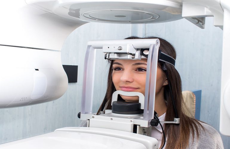
The common characteristic of cysts and similar formations that develop inside the jawbone is that they can grow for a long time without any symptoms. Most cases are noticed by chance on a film taken for another reason. This is actually good news: treatment of a formation noticed early is usually possible with more limited surgery. At the jaw cyst diagnosis stage, three-dimensional imaging reveals the true size of the formation and its relationship with neighboring structures. As DijiDent Oral and Dental Health Polyclinic, in Türkiye we consider detailed evaluation our standard approach for such findings.
What Is a Jaw Cyst?
A cyst is an enclosed formation that develops inside the bone and usually has fluid or soft tissue content. The types most frequently encountered in dentistry are root tip cysts that develop due to canal infection and developmental cysts that form around unerupted teeth. Most of these are benign; however, as they grow, they can erode the surrounding bone, push neighboring tooth roots and, rarely, reduce the strength of the jawbone.
Which Symptoms Should Be Warning Signs?
- A slowly growing, painless swelling in the jaw region
- A tooth shifting position or spacing for no reason
- Recurring gum inflammation or discharge
- A feeling of numbness in the lower lip or chin region
- A permanent tooth not erupting at the expected time
- A denture suddenly no longer fitting
The presence of these symptoms does not directly mean a cyst; however, none of them should be ignored. A feeling of numbness in particular is a finding that requires seeing a dentist without delay.
What Does Tomography Add to the Diagnosis?
On a two-dimensional film, the formation is only seen as a dark area. In three-dimensional imaging, however, the following questions can be answered: Exactly what size is the formation? Has it thinned or perforated the outer wall of the bone? Has it pushed neighboring tooth roots, or caused them to resorb? What is its relationship with the nerve canal in the lower jaw and the sinus cavity in the upper jaw? Are its borders smooth and sharp, or irregular?
These questions are important not only for surgical planning but also for gaining a preliminary idea about the character of the formation. Nevertheless, it should be clearly stated that a definitive diagnosis cannot be made by imaging alone: the final diagnosis is made by pathological examination of the removed tissue. Imaging makes it possible to establish the correct surgical plan and to avoid unnecessarily extensive intervention.
Impacted Teeth and Their Relationship with Cysts
The tissue around unerupted teeth may pave the way for cyst development over many years. This does not mean that a cyst will form around every impacted tooth; however, it is one of the main reasons why we recommend periodic check-ups for our patients with impacted teeth. Even if there are no complaints, evaluation at regular intervals allows such formations to be caught while they are small and significantly narrows the scope of the surgery needed.
How Is the Treatment Approach Determined?
Treatment is planned according to the type, size and location of the formation. In small and limited formations, removal in a single session may be possible. In large lesions, a pressure-reducing approach may be applied first and the surgery completed after the formation has shrunk. If the source of the cyst is a tooth, root canal treatment or extraction of that tooth becomes part of the plan. In all cases, the removed tissue is sent to pathology.
Why Is Follow-Up Important?
Some types of cysts have a tendency to recur. For this reason, regular check-ups and follow-up with imaging are recommended after surgery. Keeping your old images is especially valuable in this process, as it allows comparisons to be made.
Frequently Asked Questions
Most cysts seen in the jawbone are benign. Nevertheless, a definitive distinction can only be made by pathological examination; for this reason, the removed tissue is always sent for examination.
Being painless does not mean it can simply be monitored. A growing formation may require more extensive surgery over time. The decision is made according to size and course.
Bone healing is a process that takes time and varies by case. In some situations, graft support is considered.
Tooth loss is not necessary in every case. If the source tooth can be treated, the aim is to preserve it; this evaluation is made after imaging.
Some types have a possibility of recurrence. For this reason, a regular check-up program is recommended after surgery.
Do not carry a finding noticed on your film with uncertainty.
Let us carry out a detailed evaluation for swelling or numbness in the jaw region or a shadow seen on a film. Contact us to book an appointment; let us create a clear roadmap at DijiDent Oral and Dental Health Polyclinic with an examination supported by three-dimensional imaging.
This content is for general information purposes and does not replace a diagnosis. The definitive diagnosis is made by pathological examination; the treatment plan is determined by your dentist.