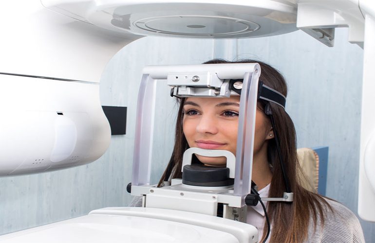
Our patients who have many crowns, metal fillings or implants in their mouths frequently ask before imaging: “Will my image come out clearly?” The source of the question is the glare and streaking that metal structures create in the tomography image. This condition, called a tomography artifact, is not a real sign of disease; it is an effect originating from the physics of imaging. As DijiDent Oral and Dental Health Polyclinic, in Türkiye we adapt the scanning protocol to the patient to reduce this effect.
Why Do Artifacts Occur?
A tomography device creates an image by measuring how much the beam passing through the tissues is attenuated. Metal structures absorb the beam much more than the surrounding tissues. When the device’s software interprets this excessive absorption, it produces dark and bright bands around the metal. This effect, seen in the image as star-shaped radiating lines, darkening or glare, can make it difficult to evaluate the neighboring region.
Which Materials Have the Greatest Effect?
- Amalgam fillings: They can create a pronounced effect due to their dense metal content.
- Metal-supported crowns and bridges: The effect increases especially in large bridges.
- Implants: A limited glare can be seen around the titanium structure.
- Orthodontic brackets and wires: They have an effect if they fall within the imaging field.
- Root canal filling materials and posts: They can make it difficult to evaluate the area around the root.
On the other hand, zirconia and all-ceramic restorations generally create less effect compared to metal-supported ones.
Do Artifacts Prevent Diagnosis?
In most cases, no. The effect is concentrated immediately around the metal; structures a few millimeters away are usually in an evaluable condition. For example, in a patient with a crown in the posterior region, bone measurement of a different region where an implant is planned can be made without any problem. The problem arises when the artifact coincides exactly with the region to be examined.
How Is an Artifact Distinguished from a Real Finding?
In an experienced evaluation, this distinction is usually not difficult. Artifacts follow a certain pattern: they radiate from the point where the metal is located, change direction in adjacent slices and do not conform to anatomical boundaries. A real pathology, on the other hand, is related to the anatomical structure, is seen consistently in adjacent slices and has definable borders. When doubt remains, your dentist may request a complementary image such as a single-tooth film or consider the clinical examination findings decisive.
What Is Done to Reduce the Effect?
The approaches we apply at our clinic are as follows:
- Narrowing the imaging field: If the metal-dense region can be left outside the field, its effect decreases.
- Adjusting the scan angle: By changing the patient’s position, efforts are made to prevent the streaking from coinciding with the region of interest.
- Selection of resolution and protocol: Device settings are adapted to the case.
- Artifact reduction algorithms: Software support available on many modern devices is activated.
- Removing removable metals: Removable dentures and accessories are removed before the scan.
- Complementary imaging: If necessary, the information is supported with an additional method such as a single-tooth film.
What Changes at the Planning Stage?
In a case where artifacts are expected, your dentist designs the imaging accordingly from the start. For example, if the region where an implant is planned is immediately adjacent to a metal-dense bridge, the imaging field can be shifted, or instead of repeating the scan with two different protocols, it can be supported with a complementary method. This preliminary assessment largely eliminates the need for a second scan.
Do I Need to Replace My Fillings?
No. Replacing a sound restoration solely for image quality is not recommended. This would be an unnecessary intervention and would cause additional loss of tooth tissue. Replacement of a restoration only comes into consideration if it has its own clinical justification.
Frequently Asked Questions
Yes. Although there is an artifact effect, evaluation can be made in most cases. The protocol is adjusted accordingly.
Unnecessary repetition is avoided. First, an evaluation is made on the existing image; however, if a critical region cannot be read, a different approach may be planned.
In an experienced evaluation, an artifact is distinguished from a real finding. In case of doubt, an additional examination may be requested.
A limited effect may be seen around the existing implant. Evaluation of different regions is usually not affected.
Fixed braces are not removed. The imaging field and protocol are planned accordingly.
The restorations in your mouth are not an obstacle to an accurate diagnosis.
Contact us to book an appointment to have your imaging needs evaluated and the protocol suitable for you determined. Let us plan the process without unnecessary repetition at DijiDent Oral and Dental Health Polyclinic.
This content is for general information purposes. The imaging protocol and evaluation are determined by your dentist according to the clinical situation.