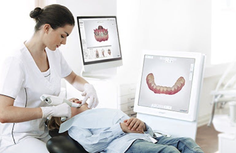
A significant proportion of dental treatments extend over months, and change occurs slowly. A person who looks in the mirror every day often cannot perceive the gradual difference in their own mouth. This is not just a matter of satisfaction; objectively documenting whether treatment is truly progressing is also clinically necessary. Thanks to the digital archive, before-after treatment comparison is now based not on impressions but on measurement. As DijiDent Oral and Dental Health Polyclinic, at our clinic in Türkiye we consider these records an integral part of treatment.
What Does a Digital Archive Include?
- The intraoral scan model taken at the start
- Interim scan records taken during treatment
- The final scan taken upon completion
- Clinical photographs and smile records
- Radiological images and design files, if any
Storing these records together allows every stage of treatment to be examined retrospectively.
How Is the Comparison Made?
Scans taken at two different times are superimposed in the software environment using common reference points. Regions that have not changed (for example, the palate surface or untreated teeth) are taken as reference. The software then visualizes the difference between the two surfaces with a color scale and expresses it numerically. In this map, shown in red-blue tones, how much change there has been in which region can be read at the millimeter level.
In Which Treatments Is It Meaningful?
In orthodontic treatment, it can be seen whether tooth movements are progressing in the planned direction. In wear monitoring, the rate of loss of tooth structure is measured. In gum treatments, the change in recession level is tracked. In implant and prosthesis applications, how the gum profile has been shaped is documented. In smile design cases, the planned result can be compared with the result achieved.
The Technical Limit of Comparison
For this method to be reliable, the reference regions must be unchanged. For example, in a case where all teeth have been restored, it becomes difficult to find a fixed reference. For this reason, comparison is mostly done regionally: an untreated area is taken as reference and the change is measured in the area of interest. Your dentist interprets the results knowing these constraints; raw numbers are not evaluated on their own.
Effect on Patient Motivation
The most common problem we encounter in long treatments is that the patient cannot feel the progress. Instead of telling a patient who has been wearing clear aligners for months “we’re doing well,” showing the superimposed image of the initial scan and the current scan is far more effective. This directly supports commitment to treatment discipline. The same applies to gum treatments and wear monitoring; seeing a measurable improvement makes it easier to maintain the recommended habits.
Documentation Value
Records are an objective basis showing the point from which treatment started. These data are valuable when getting a second opinion, changing clinics or when a different treatment is planned in the future. The initial state not being documented makes later evaluations more difficult.
Expectation Management in Aesthetic Cases
An important area of use for before-after comparison is expectation management. The goal agreed upon between the patient and the dentist at the start of treatment takes concrete form on the digital model. At the end of treatment, how close the result is to the goal is evaluated together using the same model as reference. This approach reduces the uncertainty arising from verbal agreements.
You Can Request Your Records
You have the right to access your health records. At DijiDent, scan files and clinical records are stored within the framework of the relevant legislation; they are shared digitally upon your request. We particularly recommend that our patients living abroad keep a copy of their records with them when treatment is completed.
Frequently Asked Questions
A photograph gives an image, while a scan gives a measurable model. When the two are used together, both visual and numerical comparison become possible.
The scan is taken as part of the treatment plan. You can consult your clinic for detailed information.
Yes. The initial records allow your new dentist to evaluate the process correctly and can prevent unnecessary repeat measurements.
No. Since it is an optical method, it involves no radiation and can be repeated as often as necessary.
It varies according to the type of treatment. In orthodontics they are planned at certain check-up intervals, and in follow-up cases they may be planned less frequently.
In some cases they can be digitized; however, since plaster models can wear over time, their reliability is limited.
The retention period is determined according to the relevant legislation. It is recommended that you request your own copy.
Do not guess the progress of your treatment; see it.
Contact us to book an appointment to have your initial records taken and to be followed up throughout the process. Let us measure the change together at DijiDent Oral and Dental Health Polyclinic.
This content is for general information purposes. The follow-up method and frequency are determined by your dentist according to the clinical situation.