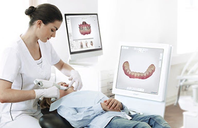
In dentistry, taking an impression is one of the invisible but most decisive steps of treatment. The fit of the crown, prosthesis or clear aligner to be made depends directly on the accuracy of the impression. An intraoral scanner performs this step with an optical scan instead of a physical mold. As DijiDent Oral and Dental Health Polyclinic, the digital impression method we use at our clinic in Türkiye aims both to increase patient comfort and to support the fit of restorations.
What Is an Intraoral Scanner?
An intraoral scanner is an optical camera with a pen-sized tip. This tip is placed in your mouth and moved slowly along the tooth surfaces. Meanwhile, the device captures hundreds of images per second. The software combines the captured images in real time and a colored three-dimensional model of your teeth appears on the screen. When the scan is complete, what you have is not a plaster model but a shareable digital file.
How Does Light Turn into Measurement?
Scanners work with different optical methods. Some project a structured light pattern onto the surface and derive depth information by measuring how this pattern is deformed on the surface. Others calculate the distance of each point using differences in focus. Whatever the method, the result is the same: every point on the surface is converted into three-dimensional coordinates, and these points combine to form the digital model.
There is an important point here: an intraoral scanner does not use radiation. It is a completely different technology from X-rays or tomography; it works only with visible light. For this reason, it can be applied in almost every situation, including pregnancy, and can be repeated as often as desired.
What Happens During the Scan?
- The tooth surfaces are dried; moisture can affect image quality.
- The scanner tip is moved in three stages: lower jaw, upper jaw and bite.
- You can also watch the model take shape on the screen.
- If there is an area that is missing, only that area is rescanned.
- The procedure is painless, requires no anesthesia and can be paused whenever you want.
Preparation Before the Scan
There are a few simple steps that increase scan quality. Plaque and soft deposits on the tooth surfaces are cleaned, because the scanner records whatever is on the surface. The cheek and tongue are kept away with special retractors or the dentist’s mirror. If there is gum bleeding, it is brought under control, because blood prevents the gum margin from being seen. This preparation takes a few minutes but directly determines the accuracy of the result.
What Is the Digital Model Used For?
The resulting model can be sent to the laboratory within seconds. Problems such as shipping time, deformation of the mold in transit or breakage of the plaster do not arise. In addition, the model can be measured on the computer: the amount of tooth wear, the level of gum recession or the difference between two scans can be compared numerically.
At DijiDent, when necessary, we combine this model with three-dimensional tomography data. Tomography shows the bone, while the scan shows the surface; when the two come together, implant and prosthesis planning can be carried out holistically.
Color Information Is Also Recorded
Most modern scanners record color information in addition to surface shape. This provides additional information to the laboratory, especially for restorations in the aesthetic zone; the color transitions and surface texture of neighboring teeth can be evaluated on the model. Having shade selection made not only by eye but with a recorded reference supports the naturalness of the result.
What Does Accuracy Depend On?
The accuracy of a digital impression depends on keeping the surface dry, the dentist’s scanning technique, the gum margin being clearly visible and the patient being able to remain still. If visibility cannot be achieved at crown margins that lie below the gum, an additional procedure may be needed to expose this area. In other words, technology alone does not guarantee the result; correct application is decisive.
Frequently Asked Questions
No. It works only with optical light and involves no ionizing radiation.
No. It is a painless procedure and requires no anesthesia. You only feel the movement of the camera tip.
It is used in many treatments; however, in some special situations a conventional impression may be preferred. The decision belongs to your dentist.
When applied correctly, comparable results are obtained. Its main advantage is comfort, speed and ease of repetition.
Yes, it is archived digitally and can be used for comparison in the future. It is also shared with you upon request.
Taking an impression is no longer a step to be feared.
If you are considering a crown, prosthesis, implant-supported solution or clear aligner treatment, you can make a comfortable start with a digital impression. Contact us to book an appointment; let us plan the process together at DijiDent Oral and Dental Health Polyclinic.
This content is for general information purposes. The impression method to be applied is determined by your dentist after the clinical examination.