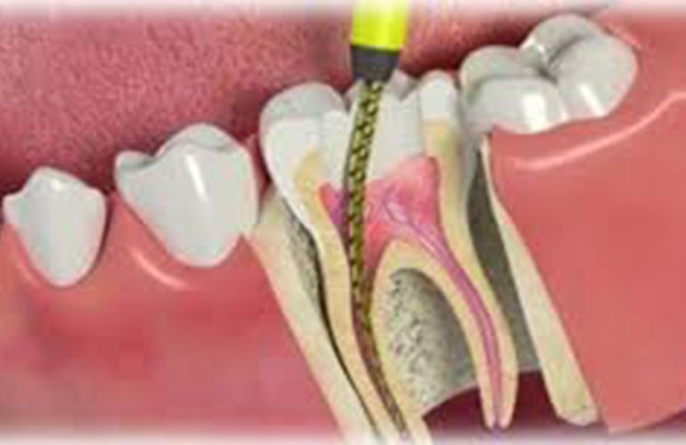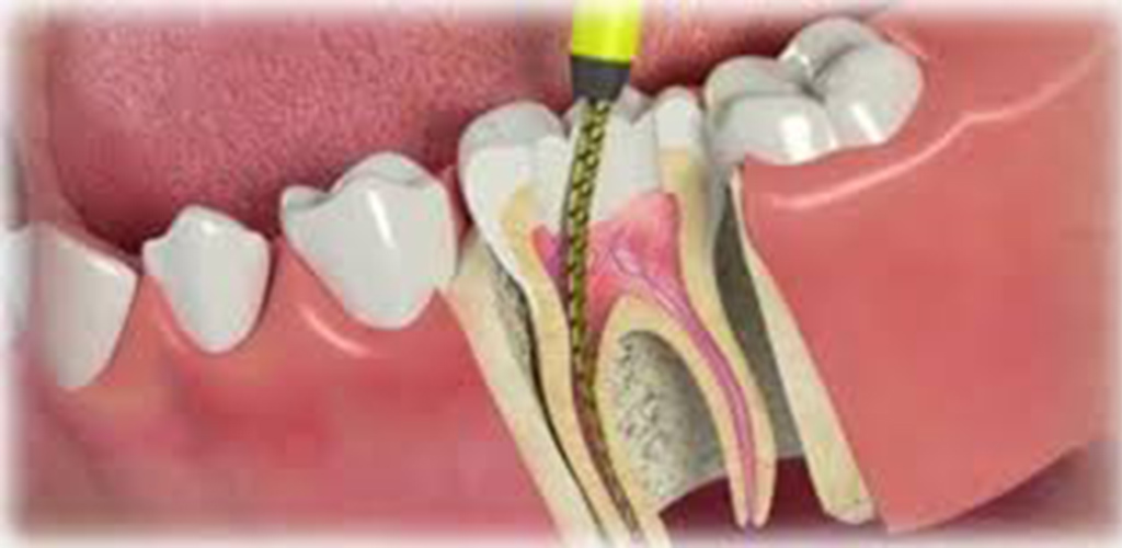
Root canals are very small and complex structures, some of which are at the limits of the human eye. Increasing vision while working on these structures can make an important contribution to the precision of the treatment. The dental operating microscope offers the dentist high magnification and strong illumination, allowing the canal system to be seen in more detail. Microscopic endodontics is a modern approach preferred especially in complex cases. As DijiDent Oral and Dental Health Polyclinic, we clearly explain to our patients in Türkiye where and how this technology makes a difference.
What Is a Dental Microscope?
A dental operating microscope is an optical device that can show the working area in the mouth much larger and directs light straight into the line of sight. Deep areas that remain in shadow with classic vision can be illuminated with the microscope and examined in detail. The microscope gives the dentist the ability to distinguish structures that are close to each other and to see very small details.
At Which Stages of Root Canal Treatment Does It Make a Difference?
Locating Canal Entrances
Additional canals, frequently found especially in upper large molars, may have very small entrances that are difficult to notice with the naked eye. The microscope makes it easier to detect these entrances. Since a missed canal can lead to the failure of the treatment later, this stage is important.
Calcified Canals
With age or after trauma, the canals may narrow or calcify. By helping to distinguish the differences in color and texture between dentin and calcified tissue, the microscope supports the canal being located safely.
Canal Retreatments
In the removal of old filling materials, pastes and posts, magnification contributes to preserving healthy tooth tissue. Seeing the remnants left on the canal walls also improves the quality of cleaning.
Managing Broken Instruments
Seeing a broken piece of instrument in the canal and removing it with special ultrasonic tips is quite difficult without magnification. The microscope is an important tool in such procedures; however, it may not be possible to remove every fragment.
Detecting Cracks and Perforations
Very fine cracks and unwanted openings on the tooth or root surface can be noticed more easily under magnification. This information helps a more accurate decision to be made about the preservability of the tooth.
Endodontic Surgery
In root-end surgeries such as apical resection, the microscope supports detailed examination of the root tip, precise work within a small surgical field and tight sealing of the root tip.
The common point of these areas of use is that more detail can be seen in small and deep structures.
What Does It Mean for the Patient?
- More detailed evaluation: The internal structure of the tooth and possible problems can be seen more clearly.
- Tissue preservation: It helps prevent more tooth tissue than necessary being removed to reach the canals.
- An option in complex cases: It may make it possible to evaluate preservation options for some teeth for which extraction is being considered.
- Documentation: Images taken with cameras connected to the microscope can be used to show the patient the condition of their tooth.
Is a Microscope Needed in Every Root Canal Treatment?
The microscope is a valuable tool; however, it is not a technology that guarantees success on its own. The success of treatment depends on many factors such as correct diagnosis, suitable cleaning and disinfection, a sealed filling and an upper restoration. In teeth with simple anatomy, magnifying loupes can also provide sufficient vision. The microscope stands out especially in difficult cases such as complex anatomy, retreatments, broken instruments, calcified canals and surgical procedures.
Visual Information for the Patient
Thanks to camera systems integrated into the microscope, images obtained during treatment can be recorded. These images can be used to show the patient a crack in their tooth, a missed canal entrance or a broken instrument fragment. Visual information helps the patient understand their situation better and participate actively in treatment decisions. In addition, the records form a valuable document if they need to be shared with another dentist in the future. At DijiDent, we keep these images as part of the patient file and share them with our patients on request.
Does the Treatment Experience Change?
Treatment done under a microscope does not feel very different to the patient from classic root canal treatment. The procedure is still done under local anesthesia and with rubber dam isolation. The patient’s head may need to be held in a certain position; for this reason, a comfortable position is provided and breaks are given if necessary. In complex cases, the session duration may be longer.
Use Together with Imaging
At DijiDent, in complex cases, we evaluate the microscope together with three-dimensional dental tomography. While tomography offers a roadmap before treatment regarding the root structure and number of canals, the microscope makes the details of this map visible during treatment. The combined use of these two tools strengthens the link between planning and application.
Frequently Asked Questions
It is root canal treatment done under a dental operating microscope that provides high magnification and illumination.
No. It is an important aid; success depends on many factors.
The microscope makes it easier to detect small canal entrances; it is especially useful in retreatments.
In complex cases the duration may be longer. In simple cases the duration is often similar.
Its necessity is evaluated according to the case. It is preferred especially in complex cases.
In complex cases, detail makes a difference.
Contact us to book an appointment to learn whether it would be beneficial to evaluate your tooth under magnification for root canal treatment. Let us create the treatment plan that suits you together at DijiDent Oral and Dental Health Polyclinic.
This content is for general information purposes and does not replace an examination by a dentist. The technology to be used and the treatment method are determined by the dentist according to the case.