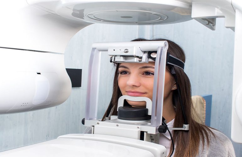
The success of implant treatment is determined not on the operating table but at the planning stage. Every step taken without knowing in advance whether the jawbone has the volume to carry the implant, where the nerve canal runs and how low the sinus floor lies is based on guesswork. 3D tomography before an implant is done precisely to eliminate this uncertainty. As DijiDent Oral and Dental Health Polyclinic, at our clinic in Istanbul we support the implant decision with measurable data.
What Does Tomography Measure in Implant Planning?
Bone Height and Width
For an implant to remain stable in the bone, it needs both sufficient height and sufficient width. On a two-dimensional film, the height of the bone can be roughly seen; however, its width cannot be seen. Yet in bone that has become thin in the lip-palate direction, if an implant of unsuitable diameter is chosen, the risk of complications increases. Three-dimensional imaging makes it possible to take this measurement at the millimeter level.
Bone Density
The hardness of the bone varies from region to region. The posterior region of the lower jaw and the posterior region of the upper jaw do not have the same density. Density information guides your dentist regarding the size of the implant, the surgical technique and the healing time.
Distance to Anatomical Structures
The nerve canal in the lower jaw, and the sinus cavity and nasal floor in the upper jaw, are the boundaries that are taken into account in implant planning. The safe distance to these structures can only be clearly determined with cross-sectional images. This is the most concrete basis of surgical safety.
From Planning to Application: The Digital Workflow
At DijiDent, we do not leave tomography data on its own. We combine the image with your digital impression taken with an intraoral scanner to create a plan in a virtual environment. In this plan, the point, angle and depth at which the implant will be placed are determined in advance. It is particularly important that the plan is compatible with the prosthetic goal: the implant should be placed not only where the bone is, but where the tooth that will go on top of it can be positioned correctly.
When necessary, a surgical guide can be produced from this plan. Placed in the mouth, this guide helps the planned position to be applied exactly during surgery. The suitability of its use is evaluated case by case.
Does the Process End If the Bone Is Insufficient?
No. One of the most valuable contributions of tomography is that it shows the extent and location of the deficiency. In this way, whether additional procedures such as bone grafting or sinus lifting are necessary and, if so, to what extent, is determined in advance. This ensures that both your treatment timeline and your cost plan are clear from the start. We aim to minimize the “surprise decision during the procedure” situation that our patients frequently experience.
What to Know Before Imaging
No special preparation is required before a tomography scan; you are not asked to fast or change your medication. You are only asked to remove items containing metal such as earrings, necklaces, glasses and removable dentures, because these can create interference in the image. It is enough to remain still for a few seconds during the scan; you do not enter a closed tunnel and the procedure is painless.
Informing your dentist about the medications you use, the surgeries you have had and your chronic conditions is also part of the planning. In particular, the use of medications that affect bone metabolism, uncontrolled diabetes and heavy smoking are factors that can change the scope of the implant decision. When we evaluate this information together with the imaging findings, we can offer you a realistic and feasible treatment plan.
The Advantage for Patients Coming from Abroad
For our patients who will be in Türkiye for a limited time, planning in advance is of critical importance. When imaging data is shared, possible scenarios can be evaluated before arrival and the session schedule can largely be created. The final decision is, of course, made after the clinical examination.
Frequently Asked Questions
In some simple cases, dentists may proceed with different methods; however, three-dimensional imaging is considered an important tool in safe planning because it shows bone volume and anatomical relationships. Your dentist makes the decision after the clinical examination.
The scan is usually completed within seconds. Including preparation and positioning, the total time does not exceed a few minutes.
Since the images appear on screen in a short time, in most cases they can be reviewed together in the same session. In complex cases, additional time may be needed for detailed planning.
The duration varies depending on the method applied and your healing rate. This information is clearly communicated to you after the tomography evaluation.
In some cases, different prosthesis options may be more suitable instead of an implant. In such a situation, the alternatives are discussed with you in detail.
Do not base your implant decision on uncertainty.
Contact us to book an appointment to find out whether your bone structure is suitable for an implant and to clarify your treatment timeline from the start. Let us carry out your digitally planned evaluation at DijiDent Oral and Dental Health Polyclinic.
This content is for general information purposes. Treatment suitability and duration vary from person to person; the decision is made after an examination by a dentist.