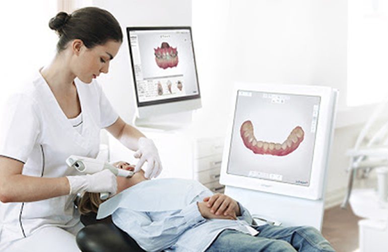
With the spread of digital scanners, there is a frequently asked question: since the device sees the inside of my mouth in such detail, can it not also detect caries? This is a reasonable expectation. However, the answer requires a correct understanding of what the technology does. Drawing a realistic framework for digital caries detection is important so that patients do not neglect check-ups out of a false sense of security. As DijiDent Oral and Dental Health Polyclinic, at our clinic in Türkiye we openly share the limits of each method.
What Does the Scanner See, and What Can It Not See?
An intraoral scanner is an optical device and records only the surface. For this reason, if there is a visible cavity, discoloration or structural defect on the tooth surface, it reflects this in the model. However, it cannot look inside the tooth. A cavity progressing beneath the enamel in the dentin layer cannot be detected by the scanner if it has not left a noticeable mark on the surface.
The Most Critical Point: Interproximal Surfaces
A significant proportion of caries begins on the interproximal surfaces where teeth are adjacent to each other. These areas cannot be seen directly either by eye or with an optical scanner, because the neighboring tooth blocks the view. Reliable evaluation of interproximal caries requires radiological methods such as bitewing films. This is not a shortcoming of the scanner; it is a matter of different tools serving different purposes.
Additional Features on Some Devices
Some scanners offer fluorescence-based imaging modes in addition to standard optical scanning. In these modes, light of a certain wavelength is sent to the tooth surface and the difference in emission between healthy tissue and demineralized tissue is evaluated. This feature can help detect early mineral loss on the surface.
However, there is a limit here too. These methods are for screening and follow-up; they are not accepted as definitive diagnostic tools on their own. In addition, tartar, staining and some restoration materials can create misleading readings. The result is always evaluated together with the clinical examination.
Caries Under Fillings
Caries developing under an existing filling cannot be seen with optical methods. Since the filling material covers the surface, the scanner records only the filling itself. Such caries are usually detected by radiological imaging or by findings at the filling margin. This is why your old fillings should be checked at regular intervals.
Methods Used in Caries Diagnosis
- Clinical examination: Visual inspection and careful use of an explorer are the basic step.
- Radiological imaging: Necessary especially for interproximal and under-filling caries.
- Optical scanning: Recording surface changes and comparing them over time.
- Fluorescence methods: Screening for early mineral loss.
- Patient history: Eating habits, sensitivity complaints and oral care routine.
None of them is sufficient on its own; a reliable diagnosis is made by evaluating them together.
Can Early Caries Be Stopped?
The situation is promising for early lesions that are confined to the enamel and have not yet formed a cavity. At this stage, it may be possible to stop or slow the process with fluoride applications, dietary adjustments and effective oral care. Once a cavity has formed, restorative treatment is needed. The value of early detection lies exactly here: a solution with less intervention.
The Value of Use for Follow-Up
When a suspicious area is detected, monitoring it at certain intervals may be preferred instead of moving directly to treatment. Digital scanning is valuable in this monitoring; by comparing records of the same area at different times, it can be evaluated whether the lesion is progressing. This approach also supports avoiding unnecessary intervention.
Frequently Asked Questions
Yes. Early caries cause no pain. Regular check-ups are the only reliable way to catch a problem while it is small.
Usually yes. Radiological evaluation may be needed for interproximal and under-filling areas.
Caries on visible surfaces can be detected by examination; however, a radiological method may be needed for a comprehensive evaluation. I do not want frequent films taken of my child. Imaging is requested only when necessary. Intervals are personalized according to risk assessment.
It is helpful for screening and follow-up; a definitive diagnosis is made with clinical and radiological evaluation.
It depends on your caries risk. Your dentist recommends a check-up interval specific to you.
Catching caries before it grows makes the treatment smaller.
Contact us to book an appointment for a comprehensive caries evaluation and a check-up program tailored to you. Let us examine your oral health holistically at DijiDent Oral and Dental Health Polyclinic.
This content is for general information purposes. The diagnostic method and treatment decision are made by your dentist after the clinical examination.